Histopathological Features:-
The gross specimen may be spherical or ovoid intact cystic masses, but often they are irregular & collapsed.
The walls vary from extremely thin to a thickness of about 5mm. The inner surface may be smooth or corrugated. The histopathological
studies shows following features ---
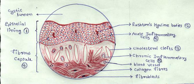
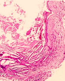
1) Epithelial Lining :- Almost all radicular
cysts are wholely or in part lined by stratified Squamous Epithelium & range in thickness from 1 to 50 cell layers. The
only exception to this is in those rare cases of periapical lesions of Maxillary Sinus. In such cases, cyst is then lined
with a pseudo stratified cilliated columnar epithelium or respiratory type of epithelium. Ortho or para keratinised linings
are very rarely seen inradicular cysts. Secretory cells or ciliated cells are frequently found in epithelial lining.
2) Rushton’s Hyaline Bodies:- In
approximately 10% of cases of radicular cysts, Rushton’s Hyaline bodies
are found in epithelial linings. Very rarely they are found in Fibrous capsule. The hyaline bodies are tiny linear or arc
shaped bodies which are amorphous in structure, eosinophillic in reaction and Brittle in nature.
3) Cholesterol Clefts:- Deposition of
Cholesterol crystals are found in many radicular cysts, slow but considerable amount of cholesterol accumulation could occur
through degeneration & disintegation of lymphocytes, plasma cells & macrophages taking part in inflammatory process,
with consequent release of Cholesterol from their walls.
4) Fibrous Capsule:- Fibrous Capsule of
Radicular Cyst is composed of mainly condensed parallel bundles of collagen fibres peripherally and a loose connective tissue
adjacent to epithelial lining.
5) Inflammatory Cells:-
Acute inflammatory cells are present when epithelium is proliferating. Chronic inflammatory cells are
present in connective tissue immediately adjacent to epithelium.
6) Mast cells, Remnants of Odontogenic Epithelium
& occasionally Satellite microcysts are also present. Some cysts are markedly vascularised.
Various kinds of Calcifications are also present.